 Image credit: AppliedSciences
Abstract
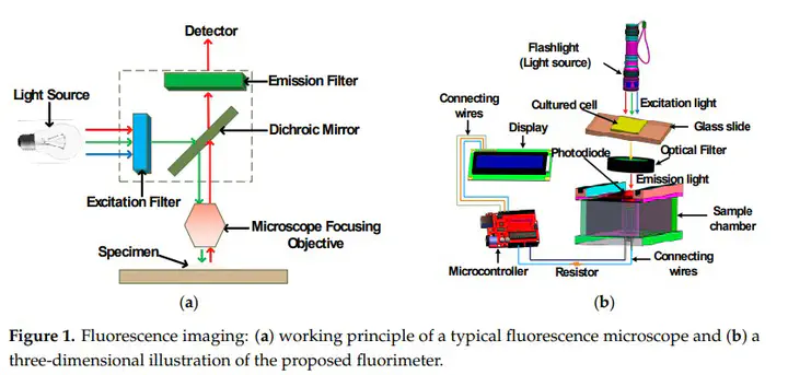
Fluorescence imaging is a well-known method for monitoring fluorescence emitted from the subject of interest and provides important insights about cell dynamics and molecules in mammalian cells. Currently, many solutions exist for measuring fluorescence, but the application methods are complex and the costs are high. This paper describes the design and development of a low-cost, smart and portable fluorimeter for the detection of colorectal cancer cell expressing IRFP702. A flashlight is used as a light source, which emits light in the visible range and acts as an excitation source, while a photodiode is used as a detector. It also uses a longpass filter to only allow the wavelength of interest to pass from the cultured cell. It eliminates the need of both the dichroic mirror and excitation filter, which makes the developed device low cost, compact and portable as well as lightweight. The custom-built sample chamber is black in color to minimize interference and is printed with a 3D printer to accommodate the detector circuitry. An established colorectal cancer cell line (human colorectal carcinoma (HCT116)) was cultured in the laboratory environment. A near-infrared fluorescent protein IRFP702 was expressed in the colorectal cancer cells that were used to test the proof-of-concept. The fluorescent cancer cells were first tested with a commercial imaging system (Odyssey® CLx) and then with the developed prototype to validate the result in a preclinical setting. The developed fluorimeter is versatile as it can also be used to detect multiple types of cancer cells by simply replacing the filters based on the fluorophore.